Fluorescent In-Situs and FCIS (Vize lab)
These pages contain protocols for performing wholemount fluorescent in situs. Instructions for two-color FISH and for a combination of fluorescent and colorimetric in situs we have called FCIS. Data can be imaged with a fluorescence stereoscope or a confocal.
FISH is not as sensitive as standard BM purple in situs, so if you have a very low abundance message it may not be detectable via fluorescence. You can however do a normal purple development for your low expression gene and a fluorescent counterstain- then overlay them to generate a FCIS image. These are both pretty and very good at highlighting quite subtle overlap
Images:
-
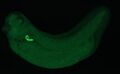
FISH, Fluorescein
-
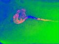
FCIS; fluorescent and colorimetric in situ
-
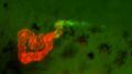
Double FISH, Fluorescein and Cy3
-
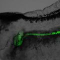
FISH plus transmitted light


Papers and other sites:
Zhou, X. and Vize, P.D. (2004). Proximo-distal specialization of epithelial transport processes within the Xenopus pronephric tubules. Developmental Biology 271: 322-338.